The skin is a very important number of organs working together in a system. It is the largest organ
in the body, and covers at least 2 m. It is a very complex structure and a vulnerable organ,
being on the outside. It is related to every other part of the body, e.g. brain, kidneys, bladder,
glands, nervous system, circulation. Any illness in these organs will show on the skin, i.e. it is a
precursor for the diagnosis of disease. Unhealthy people may lack pigment, and develop oily,
flaky, fissured and wrinkled skin. There are few curative agents for the healing of skin
conditions, because of their complexity.
If it is treated well, receives a correct balance of nutrients and sunlight,
it will provide an effective and functional barrier against most fungus, bacteria and chemical abrasion. These functions will relate further on in work practices, protection, and conditions
of humans in the horticulture environment and susceptibility to dermatitis.. To understand this
natural barrier it will be necessary to study its structure and function:
FIGURE 1.0.1 Cross Section Diagram Of The Human Skin Epidermis
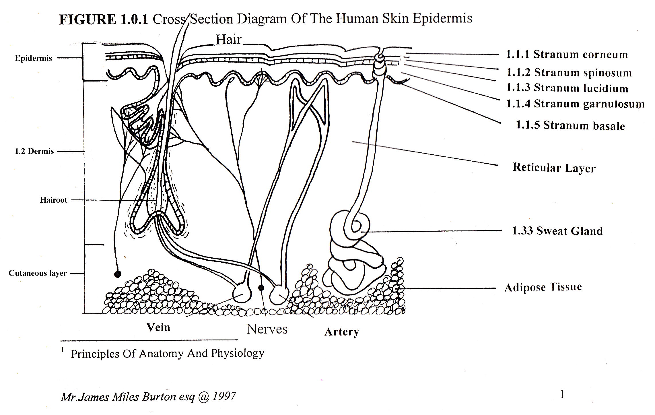
1.1 Structure
The epidermis has four basic cell types, keratinocyte, which helps to waterproof, defend and
immunise. Melanocytes, are located at the base of the skin, and protects the melanin pigments, which are responsible for the absorption of Ultraviolet light. Langerbans and Granstein cells arise from the bone marrow, and help the immune responses of the skin, and usually act as markers of antigens, which are attacked by the T-Lymphocyte immunity cells. Lipids fats, starches and proteins are also contained within these layers, aiding protection, and storage. Also embedded in the skin are blood vessels, which help to control the temperature and the nerve vessels, which with millions of nerve endings will send a barrage of information to the brain, telling it about its immediate environment. The brain is then able to respond by controlling mechanisms that will help to protect from or reduces these effects. There are four layers in the skin, five in the soles and palms:
1.1.1 Stranum corneum: The top layer consists of 25 layers of dead cells, filled with tough keratin the substance that makes nails, and used in protective chainsaw suits. These are continuously being shed and replaced. It serves as an effective barrier against light, heat, bacteria and chemicals. In the manufacture of these cells, a process called keratinisation, new cells are pushed up from the basal layers. The period between forming and shedding takes about two weeks.
1.1.2 Stranum spinosum: This layer contains many sided cells that fit together.
1.1.3 Stranum lucidium: This layer is found only in thick skin, such as the palm and soles.
They contain clear dead cells called eleidin, which is eventually transformed into keratin.
1.1.4 Stranum garnulosum: The second layer consists of 4 rows of flattened cells that contain forms of stained keratin. This provides a waterproofing protein. They are in a vigorous state of degradation.
1.1.5 Stranum basale: This single layer pushes up towards the surface. The nuclei disintegrate and
become the next layer. Other cells may arise and forms hair follicles or glands.
1.2 Dermis the second part of the skin. It is composed of many connective tissues containing fibres.
It is very thick in parts, especially the buttocks. It has nerves, blood vessels, and glands imbedded in it.
The papillary region consists of loose connective tissues, which contain elastic fibres, these project into the epidermis, and are responsible for fingerprints. It also allows the whole skin to move freely over the underlying structures. Fat deposits also occur here. The remaining lower portion of the skin is called the reticular layer,
and consists of dense connective tissues with interlacing elastic tissues. The spaces in-between are filled by epidermal derivatives. The region is connected to the underlying organs and bones by the subcutaneous layer.
1. 3 Epidermal derivatives: These structures develop from the division and specialisation of cells in
the subcutaneous layer, and provide an important role in protection and temperature regulation. Hairs growths are distributed over the body to provide protection from ultra violet light, and particles. They are also sensitive to touch and respond to emotions. It consists of a shaft and a root /bulb. The root connects with blood vessels, which provide it with cells that specialise, divide and push the older cells upwards which then die. The smooth muscle surrounding the root are called the arrector pillii, which contracts under stress cold fright for extra protection causing goose bumps.
1.3.1 Glands Three Types: The secreting portions lie in the dermis, and open into the necks of the hair follicles, those surrounding the penis, eyelids lips and anus open directly onto the skin surface. They vary in size and are largest in the neck and chest. They do not occur in the soles and palms. They secrete an milky oily substance called sebum, a mix of fats, cholesterol, proteins and salts. It prevents the hairs from drying out, prevents excessive evaporation of water, keeps it soft and pliable and inhibits the growth of bacteria.
1.3.2 Sweat Glands: Approcine sweat glands are branched tubular organs. They are limited to the
pubic regions, breasts and axilla. The secretory portion is located in the dermis, and begins to
function at puberty. They excrete a milky perspiration, that may be related to copulation.
1.3.3 Eccrine Sweat Glands: are much more common, and are simple coiled structures located in the subcutaneous layer. The excretory portions are located in the epidermis. The secretory portion here is more watery. Perspiration, the substance produced by these glands contain a mixture of ammonia, lactic ascorbic acids, salts and urea. The principle function is to control the temperature of the body by evaporation of sweat. Secondly to excrete wastes, and third to inhibit the growth of bacteria fungi and protect the upper surface. However an excess of perspiration may aid the toxic effects of chemicals on the skin.
1.4 Functions Of The Skin
1.4.1 Homeostasis Of Body Functions: Mammals are warm blooded and need to maintain an internal body temperature of 37C in order to function correctly. It responds to heat as a stress.Thermoreceptors pick up the stimulus, and send an impulse to the brain, which then signals the sweat glands to perspire. As this evaporates, the body is cooled. 22% of heat is lost in this way. The skin will loose up to 1 gallon of water every hour under strenuous activity and work.
1.4.2 The Skin And Immunity The skin provides an effective natural physical barrier to disease, abrasion, radiation and bacteria, because of toughened keratin cells, fat within, hairs on the surface, glands which secrete defensive, lubricating, anti bacterial acids, alkalis, ammonias and water. Also, within the skin, are immune cells. An antigen presenting itself to the skin will bind to Langerhans and Granstein cells, acting as a marker for the activation of T-Lymphocytes cells, which destroy it. The skin, if kept healthy from inside and out, by stress free living and correct food consumption, will defend adequately and heal well, it will not need extra medicaments, which often hinder this natural process.
1.4.3 Reception Of Stimuli The skin contains millions of receptors in the nerve endings, and respond to stress, pressure or abrasion by sending increasingly numerous signals to the brain, which is then able to respond mechanically by removing the body from that harmful environment or object, so aiding in the immune response.
1.4.4 Synthesis of Vitamin D Is facilitated by the precursor molecules in the subcutaneous layer, when exposed to ultraviolet light, producing Vitamin D, This is then transported into the blood for use in protein synthesis. It is protected by the melanocytes, and gives the skin its distinctive pinkish colour. Persons of
African origin have a negroid colour, because these cells have over synthesised the production of specialised protective melanin pigments and melanocytes.
1.5 Ageing of the skin
At birth the skin has not developed a sufficient protective layer, or facilitated the synthesis of immune cells.
The skin is often seen to be transparent, and therefore is sensitive to damage, and must be protected by extra clothing. At puberty, glandular, hair development and immune systems begin to function at an increased rate, giving extra protection against the adult world. During puberty, the skin is very active and vulnerable to sensitisation by allergens. At age 20 however the skin begins to deteriorate, and by the age of 50 is in a rapid state of degradation. Collagen fibres begin to fall apart, elastic fibres stiffen, and thicken into lumps. This wrinkles the skin. Oil glands ceases production, and melanin production decreases, leading to pallid colour, and grey hairs. The keratin cells also cease production and so become thin and stiff. It heals poorly, after wounding, and becomes susceptible to disease, cancer, shingles and dermatitis. Exposure to ultra violet light will accelerate ageing.
Dermatitis In the Horticulture Industry By James M. Burton In Association With Pencoed College Copyright 1997.